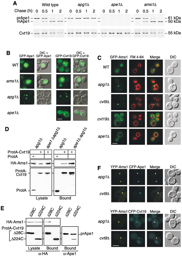
Figure 5. Precursor Ape1 Is Required for the Efficient Transport of Cvt19 and the Cargo Protein Ams1.
(A) Wild-type, apg1Δ, ape1Δ, and ams1Δ cells were pulse labeled for 10 min in SMD medium and chased for the indicated times in SMD medium. Ape1 and Cvt19 were immunoprecipitated from cell extracts and analyzed by SDS-PAGE.
(B) Localization of GFP-Ape1 or GFP-Cvt19 in wild-type, ams1Δ, apg1Δ, and ape1Δ strains by fluorescence and DIC microscopy. Bar, 5 µm.
(C) Localization of GFP-Ams1 in wild-type, apg1Δ, cvt9Δ, cvt19Δ, and ape1Δ strains by fluorescence and DIC microscopy. Cells were grown to midlog phase and labeled with FM 4-64 in SCD medium. Bar, 5 µm.
(D) Ams1 associates with Cvt19, independent of Ape1. The apg1Δ or ape1Δ apg1Δ cells were transformed with pHA-Ams1 and pRS416-CuProtA or pCuProtA-Cvt19. IgG Sepharose was used to precipitate ProtA-Cvt19 from cell lysates. The left and right panels show the protein blots of total cell lysates (Lysate) and the IgG precipitates (Bound), respectively, which were probed with anti-HA antibody.
(E) The Ams1 binding site in Cvt19 is different from the prApe1 binding site. Total lysates from apg1Δ cvt19Δ cells expressing HA-Ams1 and either ProtA-Cvt19Δ28C or ProtA-Cvt19Δ224C were used to precipitate ProtA-Cvt19 proteins with IgG Sepharose. Total cell lysates (Lysate) and the IgG precipitates (Bound) were analyzed by Western blot with anti-HA or anti-Ape1 antisera.
(F) Colocalization of YFP-Ams1 with CFP-Ape1 or CFP-Cvt19 in apg1Δ and cvt9Δ cells by fluorescence and DIC microscopy. Bar, 5 µm.

