Figure 1.

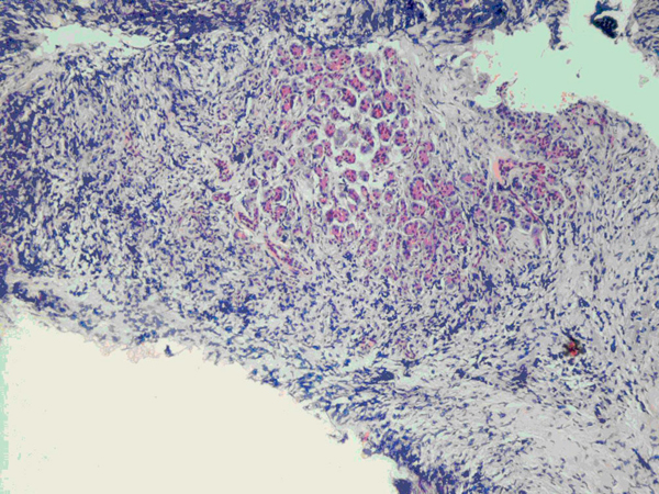
Low-power view of pancreatic biopsy showing residual pancreatic acini centrally, surrounded by a dense infiltrate of smaller cells (hematoxylin and eosin stain, ×100).

Low-power view of pancreatic biopsy showing residual pancreatic acini centrally, surrounded by a dense infiltrate of smaller cells (hematoxylin and eosin stain, ×100).