
Figure 4.
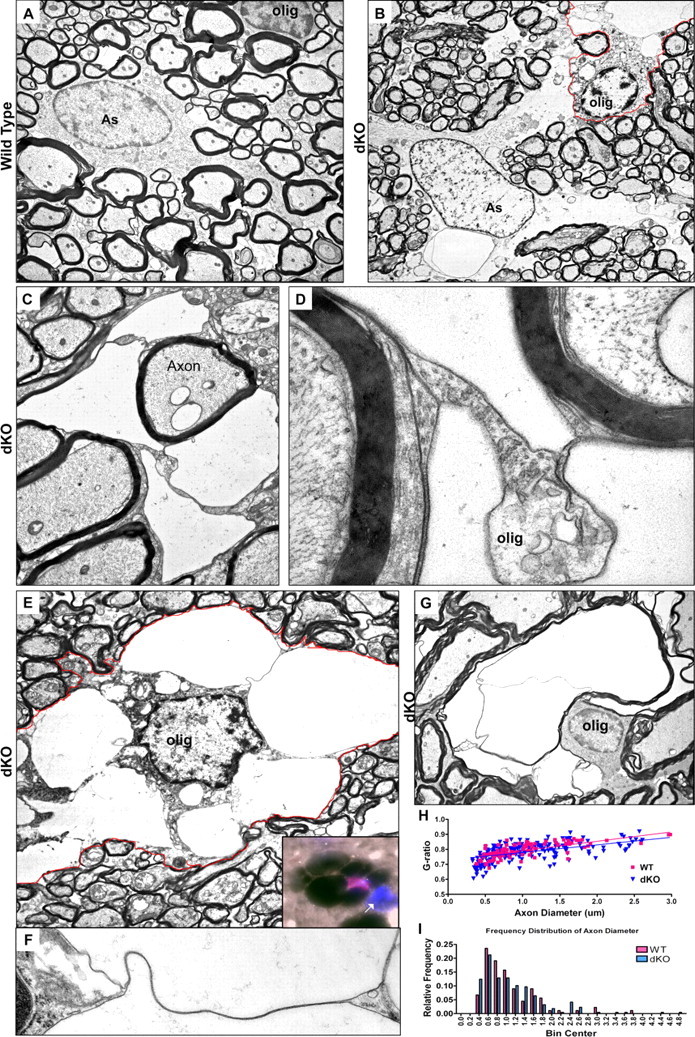
Electron micrographs depicting oligodendrocyte vacuolation within dKO white matter. A, Astrocyte (As) and oligodendrocyte (olig) in white matter of wild-type spinal cord. The astrocyte can be identified by marginal chromatin and cytoplasmic intermediate filaments. B–G, Vacuolated oligodendrocytes and astrocyte in dKO white matter. B, Vacuolated oligodendrocyte abutting edematous astrocyte in dKO cerebellar white matter. The oligodendrocyte is identified by marginal chromatin, microtubules, and relatively electron-dense cytoplasm. Note that the astrocyte also shows extensive pathology with loss of well defined cytoplasmic organelles. The oligodendrocyte is outlined in red. C, Highly compartmentalized oligodendrocyte cytoplasm. D, Higher-power view of the field in C helps identify the compartmentalized cytoplasm as oligodendrocytic due to the presence of microtubules and lack of filaments, and because its membranes are contiguous with compact myelin. E, An oligodendrocyte containing numerous membrane-bound vacuoles within its cytoplasm. The oligodendrocyte is outlined in red. Inset: immunohistochemical staining showing the nucleus of a vacuolated cell positive for both DAPI (blue) and olig2 (red). Note the unaffected, olig2-negative nucleus at right (arrow). F, Detail from E showing a continuous ribbon of oligodendrocyte cytoplasm connecting the perinuclear cytoplasm and plasma membrane between two vacuoles, demonstrating that the vacuoles are contained within the oligodendrocyte. G, Intramyelinic edema. H, I, dKO and wild-type g-ratios (H) and axon diameter (I) are not significantly different in the cerebellum. Magnification: (A), 4000×; (B), 2600×; (C), 6000×; (D), 35,000×; (E), 5000×; (F), 21,000×; (G), 3600×.