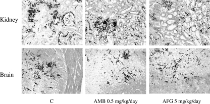
FIG. 3.
Histopathological sections of kidney and brain tissues stained with Grocott Gomori (original magnification, ×25) from mice infected with 3.2 × 10⁶ conidia of the A. fumigatus F3 isolate. Representative histopathological sections of kidney and brain tissues from control mice (C) and from mice treated for three consecutive days with AMB at 0.5 mg/kg/day and AFG at 5 mg/kg/day are shown.

