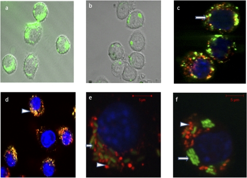
FIG. 2.
Confocal microscopy. (a and b) Uptake of N2F (a) and N1F (b) nanostructures into J774A.1 cells. (c and d) Colocalization of nanostructures with endosome/lysosome after incubation for 2 h. Subcellular colocalization of N1F (arrow) (c) and N2F (arrowhead) (d) nanostructures is shown by yellow-to-orange spots formed by green nanoparticles and red endosomes/lysosomes, showing that a majority of the N2F hydrophilic nanostructures appear to reside in endosomes. (f and e) Salmonella (arrow)-infected J774A.1 macrophage cells incubated for 4 h with N1R and N2R nanostructures (arrowheads).

