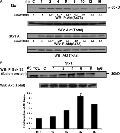
FIG. 5.
Stx1 and Stx1A− activate Akt in THP-1 cells. (A) THP-1 cells were stimulated with Stx1 or Stx1A− for 1 to 16 h. At each time point, cell lysates were prepared and Western blotting (WB) performed using antibodies specific for phospho-Akt (S473). Blots were stripped and reprobed with antibodies recognizing total Akt for equal protein loading (bottom panel). C, lysates prepared from control cells maintained for 16 h without Stx1. Band intensities were measured by densitometry, and n-fold changes are expressed beneath each lane. (B) THP-1 cells were treated with Stx1 for 1 to 8 h. At each time point, cell lysates were prepared. Supernatants collected from the lysates were precleared and incubated with an anti-Akt monoclonal antibody. An Akt kinase assay was performed with the resultant immunoprecipitates using a synthetic GSK-3β fusion protein substrate in the presence of ATP. Proteins were then separated by SDS-PAGE and Western blotting performed using an antibody recognizing phospho-GSK-3β. A representative Western blot is shown in the upper panel. TCL, total cellular lysate; C, lysates from control (unstimulated) cells; IgG, negative control. The bar graph shown in the lower panel is the n-fold-induction of Akt kinase activity following Stx1 treatment of THP-1 cells derived from at least three independent experiments. Asterisks (*), significant difference (P < 0.05) for toxin treatment results versus those with control cells.

