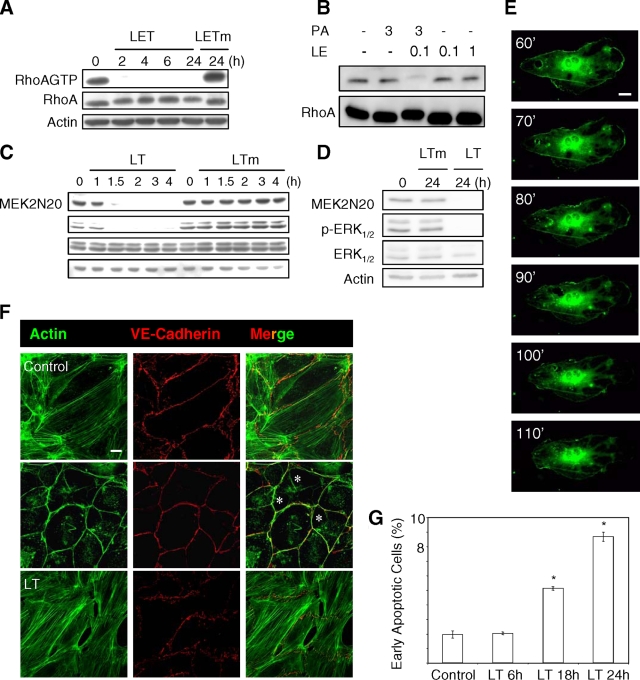
FIG. 2.
PA-dependent delivery of LE chimera into endothelial cells. HUVEC monolayers were intoxicated with LET (3 μg/ml PA plus 1 μg/ml LF1-254-EDIN), LETm (3 μg/ml PA plus 1 μg/ml LF1-254-EDINR185E), LT (3 μg/ml PA plus 1 μg/ml LF), or LTm (3 μg/ml PA plus 1 μg/ml LFE687A) for the indicated periods of time. (A and B) The levels of active RhoA (labeled RhoA-GTP) were determined by using glutathione S-transferase-Rhotekin receptor-binding domain pull down. The cellular level of RhoA was determined by anti-RhoA immunoblotting of total protein extracts. The Western blot of anti-RhoA and/or anti-actin shows equal protein loading. (C and D) HUVEC monolayers were intoxicated, and the effect of LF was assessed by anti-MEK2N20 and anti-phospho-ERK1/2 immunoblotting. The Western blot of anti-actin and anti-ERK1/2 shows equal protein loading. (E) Formation of macroapertures in HUVEC monolayers transfected with green fluorescent protein-actin and treated with LET (images from Video S1 in the supplemental material; intervals in minutes). (F) Actin cytoskeleton organization in control or intoxicated monolayers for 2 h with LET or for 24 h with LT. F-actin was labeled with FITC-phalloidin (green), and VE-cadherin adhesion molecules were labeled with anti-cadherin 5 (red). The asterisks indicate LET-induced macroapertures. Scale bar, 10 μm. (G) Effect of LT on endothelial cell apoptosis. Cells were treated with medium alone or with medium supplemented with LT. The bars and error bars indicate the means ± standard deviations of the percentages of early apoptotic cells (phycoerythrin-annexin V positive and 7-amino-actinomycin D negative) for three independent experiments. Statistical data analysis was performed with unpaired two-tailed t tests (*, P < 0.01 compared with control cells).

