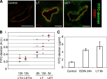
FIG. 4.
LET-induced permeability in animals. Mice were inoculated intravenously with LT (100 μg PA plus 100 μg LF), LTm (100 μg PA plus 100 μg LFE687A), LET (100 μg PA plus 100 μg LF1-254-EDIN), LETm (100 μg PA plus 100 μg LF1-254-EDINR185E), or EDIN (1 μg). (A) Six hours following toxin injection mice were inoculated with FITC-dextran (50 μg/mice), and they were euthanized 30 min later for analysis. The images are representative photomicrographs of FITC-dextran (green) diffusion in lung vessels corresponding to a confocal optical section. Anti-CD31 revealed endothelial cells (red). Scale bar, 50 μm. (B and C) Quantitative measurement of pulmonary vessel permeability by diffusion of FITC-dextran in the lung. At the indicated times mice were inoculated with FITC-dextran (50 μg/mouse) 30 min prior to recovery of FITC-dextran in lung as described in Materials and Methods. A.U., arbitrary units. (B) Circles indicate values obtained for individual mice. (C) Pulmonary endothelium permeability determined for eight mice per condition (means ± standard deviations).

