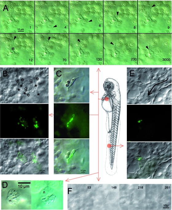
FIG. 2.
In vivo microscopic observation of early Listeria infection. Larvae at 55 hpf were examined either in the region of the duct of Cuvier (A, B, C, D, and F) or in the CHT (E); the central scheme depicts the location but not the orientation. Observations were performed 1 to 2 h following i.v. injection of GFP-expressing L. monocytogenes (A, B, C, E, and F) or of L. innocua (D). (A) Phagocytosis of blood-borne L. monocytogenes by a macrophage in the duct of Cuvier. Ten frames were extracted from a 1-h DIC movie. The number on lower right corner of each frame indicates time in seconds; time zero starts at around 1 hpi. The position of the bacterium is indicated by the arrowhead, and the nucleus of the macrophage is indicated by an n. Other cells are erythrocytes temporarily trapped due to the pressure of the coverslip on the yolk sac. (B, C, and E) Bacterium-containing cells viewed by DIC (top), fluorescence (middle), or both (bottom). (B) Typical aggregation of cells observed in the duct of Cuvier at 1 hpi, with bacterium-laden macrophages (asterisks), granulocytes that do not appear to contain bacteria (g), and erythrocytes (e). (C) Isolated macrophage observed in the duct of Cuvier, with several internalized bacteria. n indicates its nucleus; a freshly divided pair of erythrocytes (e) is transiently sticking to it. (D) In larvae infected with L. innocua, 1 or 2 h after infection, bacteria are found gathered inside a large, single phagosome per macrophage (DIC optics). n indicates the nucleus. (E) Bacterium-containing myeloid cell observed in the CHT near a caudal vein segment where circulating erythrocytes are seen (the direction of flow is indicated by the curved arrow). (F) A few bacteria stick to the endothelium or the yolk cell surface as shown here and can divide extracellularly. The time in minutes postinfection is indicated on each frame.

