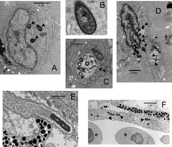
FIG. 3.
Ultrastructural analysis of L. monocytogenes infection of zebrafish larvae. (A to D) Standard staining at 3 hpi. (E and F) Tilney staining at 24 hpi. (A) Macrophage containing three intracytoplasmic bacteria (arrowheads). (B) Close-up of one of these bacteria showing the absence of any membrane between the bacterial cell wall and cytosol. (C) Macrophage with a large phagosome containing bacteria at various stages of degradation. The dark object in the cytosol, indicated by an arrow, does not harbor a cell wall and is probably a fat globule. (D) Neutrophilic granulocyte, identifiable by its typical spindle-shaped granules (arrows), with at least two killed bacteria (bottom arrowheads) within a phagosome and a third one probably still alive (top arrowhead) in another phagosome. (E) Typical cell protrusion containing a dividing bacterium propelled by an actin tail. (F) Infected endothelial cell, with some bacteria indicated by arrowheads and vessel lumen on the bottom of the image. n, nucleus; m, melanophore; e, erythrocyte.

