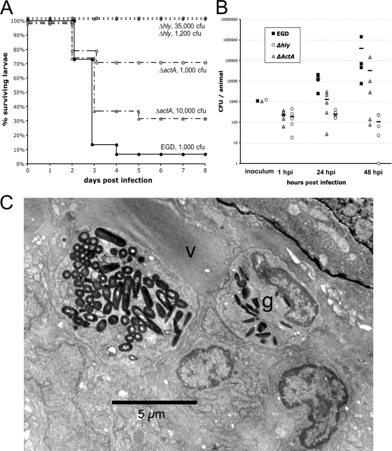
FIG. 4.
Listeriolysin and ActA are virulence factors for L. monocytogenes in the zebrafish model. (A) Survival curve of zebrafish larvae infected i.v. with wild-type L. monocytogenes (EGD) or with ΔactA or Δhly EGD mutants at various doses and incubated at 28°C (n = 15 to 24 per group). (B) Enumeration of bacteria inside whole larvae at various time points following i.v. infection. Load was not measured at 1 hpi in EGD-infected larvae in this experiment (see Fig. 1C for a similar measurement). At days 1 and 2, CFU numbers were statistically different when Δhly mutant- and EGD-injected larvae were compared (P < 10−3) or, less significantly, when ΔactA mutant- and EGD-injected larvae were compared (P < 0.05). (C) TEM image of a typical macrophage in a zebrafish infected with the Δhly mutant at 3 hpi, with a huge phagosome containing many bacteria. The nucleus is not visible in this plane. The macrophage is in direct contact with the lumen of the vein (v). A bacterium-free neutrophilic granulocyte (g) is also visible in its vicinity.

