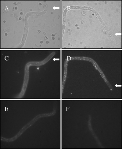
FIG. 5.
Localization of CFH and C4BP bound on Loa loa MF. The MF were enriched from EDTA-blood by the Percoll gradient method, washed, and subjected to immunofluorescence staining using polyclonal goat anti-CFH (C) or rabbit anti-C4BP (D) antibody and Alexa 488-conjugated secondary antibodies. The photographs were taken from representative MF using bright-field (A and B) or fluorescence (C and D) microscopy. Control photographs with only the secondary antibodies are shown in panels E and F. The typical extended sheath in the anterior end of Loa loa MF is indicated with a arrows in panels A, B, C, and D.

