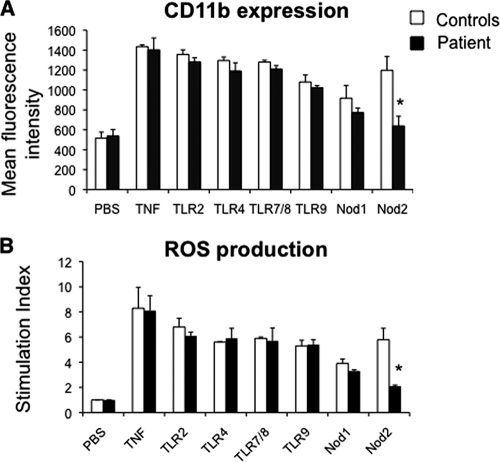
FIG. 2.
Impaired PMN functions after Nod2 stimulation in a patient with granulomatous mastitis due to C. kroppenstedtii. (A) CD11b expression at the PMN surface. Whole-blood samples were incubated at 37°C for 45 min with phosphate-buffered saline (PBS), TNF (100 U/ml), TLR agonist macrophage-activating lipopeptide 2 (10 ng/ml) (TLR2/6), lipopolysaccharide (10 ng/ml) (TLR4), R-848 (10 μg/ml) (TLR7/8), or CpG DNA (100 μg/ml) (TLR9), the Nod1 agonist C12-iE-DAP (lauroyl-γ-d-glutamyl-meso-diaminopimelic acid) (50 μg/ml), or the Nod2 agonist MDP (10 μM). Samples were then stained with phycoerythrin-conjugated anti-CD11b monoclonal antibody at 4°C for 30 min. Results are expressed as mean fluorescence intensity. (B) PMN oxidative burst. Whole-blood samples were pretreated with hydroethidine (1,500 ng/ml) for 15 min at 37°C and then incubated for 45 min with PBS, TNF, or TLR and Nod agonists as described above, followed by fMLP stimulation (10−6 M, 5 min). Results are expressed as a stimulation index (ratio of the mean fluorescence intensity of stimulated cells to that of unstimulated cells). Values that are significantly different (P < 0.05) from those of the controls are indicated by an asterisk. Data are reported as means plus standard errors of the means (error bars). Comparisons were based on analysis of variance and Tukey's posthoc test, using Prism 3.0 software.

