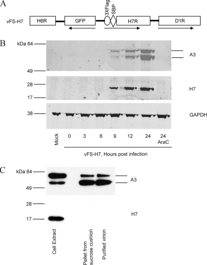
FIG. 2.
Expression of H7. (A) Schematic representation of a portion of the genome of recombinant VACV vFS-H7. ORFs are indicated by boxes, and the directions of transcription are indicated by arrows. The 3×Flag and streptavidin-binding peptide (SBP) tags are fused to the N terminus of H7. (B) Expression of H7. BS-C-1 cells were mock infected or infected with vFS-H7 in the absence or presence of AraC and harvested at indicated times after infection. Whole-cell lysates were analyzed by SDS-PAGE, and the proteins were transferred to a membrane and probed with anti-Flag M2 mouse MAb to detect H7 and a rabbit polyclonal antibody to A3, followed by mouse- and rabbit-specific fluorescent secondary antibodies. Fluorescent images are shown. The blot was then stripped and reprobed with antibody to glyceraldehyde-3-phosphate dehydrogenase (GAPDH) as a loading control. (C) Analysis of purified MVs. Cells infected with vFS-H7 were lysed, and MVs were purified by sedimentation twice through sucrose cushions and a sucrose gradient. Fractions of the cell lysate, pellet from the second sucrose cushion, and purified virions were analyzed by SDS-PAGE and Western blotting using antibodies to the Flag epitope to detect H7 and specific antibody to A3. The bands were detected by chemiluminescence.

