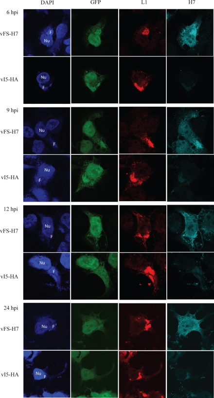
FIG. 3.
Localization of H7 in infected cells by confocal microscopy. HeLa cells grown on coverslips were infected with either vFS-H7 or control vI5-HA virus (35). The vI5-HA virus was chosen as a control because it shows no defect in replication and expresses GFP. At 6, 9, 12, and 24 h, the cells were fixed and permeabilized. L1 and H7 were detected with a mouse MAb and rabbit polyclonal antibody, respectively, followed by species-specific fluorescent secondary antibodies. DNA was stained with DAPI. Nu, nucleus; F, viral factory; hpi, hours postinfection.

