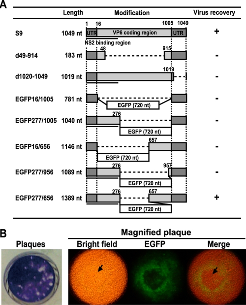
FIG. 4.
Mapping the sequences of the S9 transcripts essential for virus rescue. (A) Schematic representation of modified S9 transcripts. The NS2 binding region (nt 1 to 275) is shown as black bars. BSR-VP6 cells were transfected with five BTV T7 transcripts (S1, S3, S4, S6, and S8), followed by a second transfection 18 h later with uncapped modified S9 transcripts, together with nine other capped transcripts. Each transcript was at 50 ng per transfection. Virus recovery is shown as a plus in the right column. (B) Plaque formation and the expression of EGFP were observed in BSR-VP6 cells pretransfected with five capped transcripts (S1, S3, S4, S6, and S8), followed by a second transfection with uncapped chimeric S9 EGFP277/656, together with the nine remaining capped transcripts, using a fluorescence microscope. Upon observation of EGFP expression, the cells were stained with 0.2% (wt/vol) crystal violet (left). Cytopathic effect is clearly observed in a magnified plaque (indicated with arrows).

