
FIG. 6.
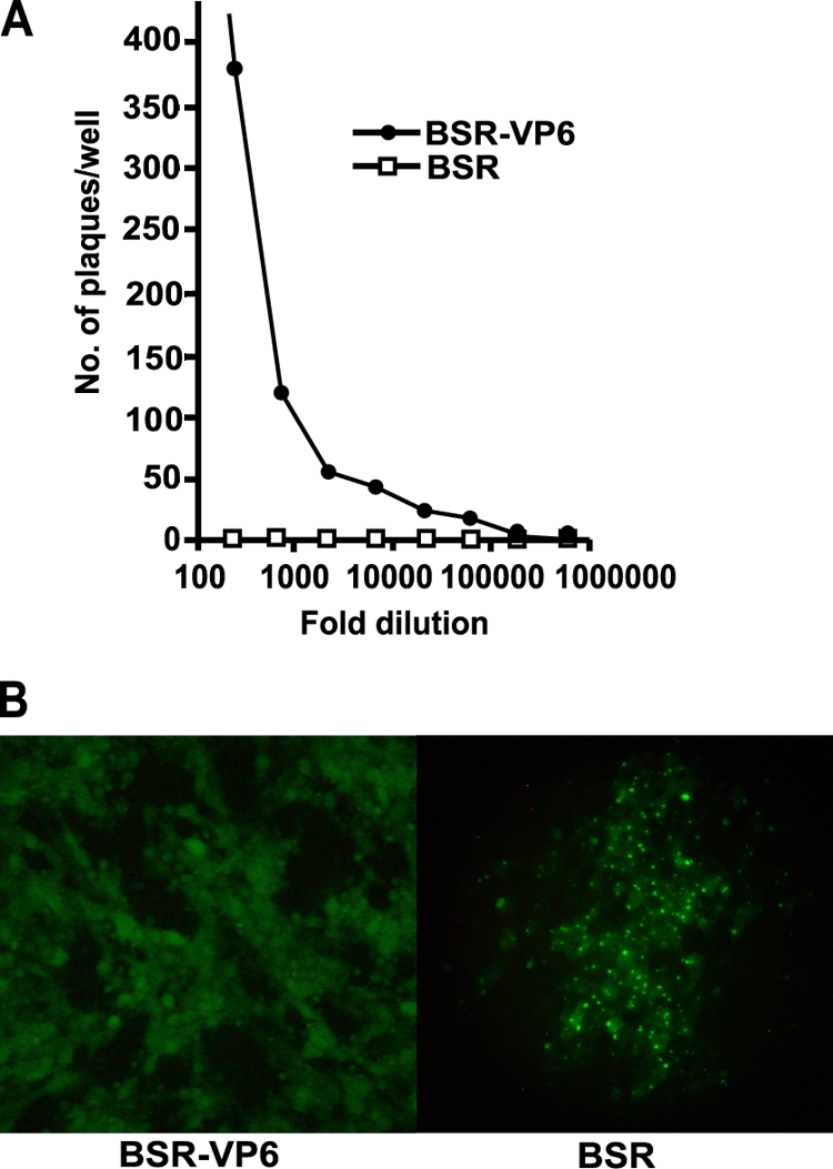
Infectivity assay of BTVS9EGFP. (A) Serial-dilution analysis of BTVS9EGFP. BSR-VP6 cells and BSR cells were infected with BTVS9EGFP stepwise diluted at one in three. The plaques were counted 2 days postinfection. (B) Expression of EGFP was observed in BSR-VP6 cells and BSR cells 2 days postinfection using a fluorescence microscope. (Left) BSR-VP6 cells infected with BTVS9EGFP. (Right) BSR cells infected with BTVS9EGFP. Note that compared to the BSR-VP6 cells, the BSR cells showed EGFP as distinct punctae.