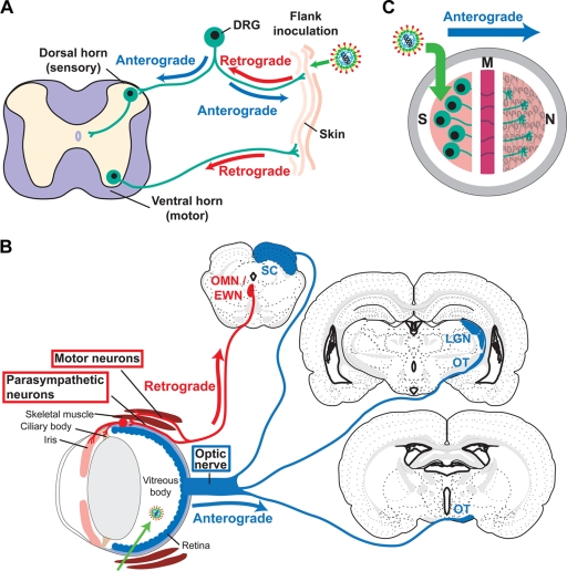
FIG. 1.
Model systems to study HSV-1 neuronal spread. (A) Mouse flank model. Virus was scratch inoculated onto the skin, where it replicates, spreads to innervating neurons, and travels in a retrograde direction to the neuron cell body in the DRG. After replicating in the DRG, the virus travels in an anterograde direction back to the skin and into the dorsal horn of the spinal cord. Motor neurons also innervate the skin, allowing virus to reach the ventral horn of the spinal cord by retrograde transport. (B) Mouse retina model. Virus is injected into the vitreous body, from which it infects the retina as well as other structures of the eye, including the ciliary body, iris, and skeletal muscles of the orbit. From the retina, the virus is transported into the optic nerve and optic tract (OT) (anterograde direction) and then to the brain along visual pathways. Anterograde spread is detected in the lateral geniculate nucleus (LGN) and superior colliculus (SC). From the infected ciliary body, iris, and skeletal muscle, the virus spreads in a retrograde direction along motor and parasympathetic neurons and is detected in the oculomotor and Edinger-Westphal nuclei (OMN/EWN). Only first-order sites of spread to the brain are indicated. (Brain images were modified and reproduced from reference 47 with permission from of the publisher. Copyright Elsevier 1992.) (C) Campenot chamber system. Campenot chambers consist of a Teflon ring that divides the culture into three separate compartments. Neurons are seeded into the S chamber and extend their axons into the M and N chambers. Vero cells are seeded into the N chamber 1 day before infection. Virus is added to the S chamber and detected in the N chamber, a measure of anterograde spread.

