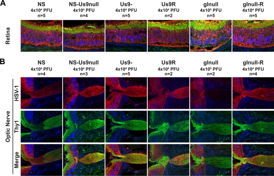
FIG. 4.
Anterograde transport into the optic nerve. Mice were infected in the vitreous body with WT, mutant, or rescue strain HSV-1, and tissues were harvested 5 dpi. Sections were stained for immunofluorescence with anti-HSV-1 (red), the neuronal marker anti-Thy1 (green), and DAPI (blue). (A) Ganglion cell neurons form the innermost layer of the retina, which is shown in green at the top of each image. Magnification, ×200. (B) The retina is shown on the left side of each image, while the optic nerve is shown on the right, at the site of exit from the retina. Magnification, ×100.

