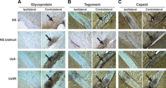
FIG. 6.
Glycoprotein, tegument, and capsid antigens are transported into the optic tract. Mice were infected in the vitreous body with 4 × 10⁴ PFU HSV-1 NS, NS-Us9null, Us9-, or Us9R, and tissues were harvested 5 dpi. Optic tracts ipsilateral and contralateral to the injected eye are shown at ×200 magnification. Antigen staining in the contralateral optic tract is indicated with black arrows. Brains were stained with the following antibodies against glycoprotein, tegument, or capsid: R122 anti-gD (NS, NS-Us9null) or R118 anti-gC (Us9-, Us9R) (A); PSU74 anti-VP22 (B); and NC-L anti-capsid (C). The antibodies against VP22 and light capsid produced higher background staining levels than did the antibodies against gD and gC.

