FIG. 7.
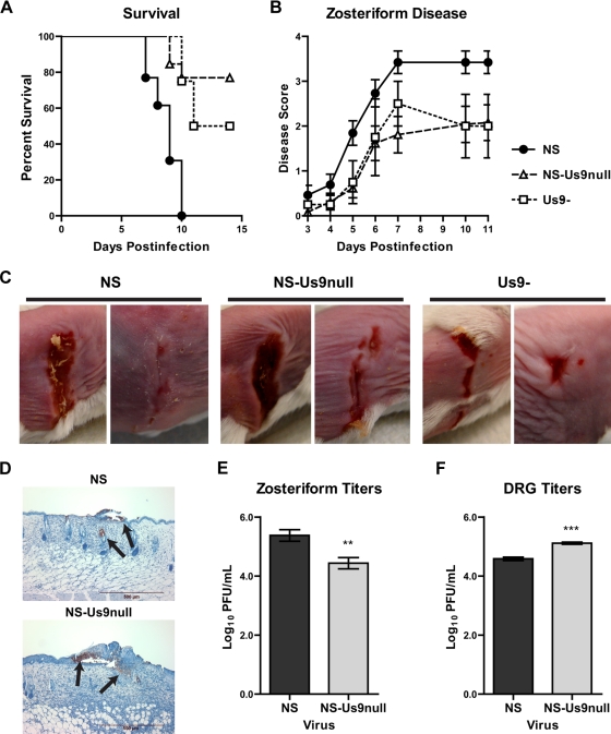
Zosteriform disease in the mouse flank model. Mice were scratch inoculated with 5 × 10⁵ PFU NS, NS-Us9null, or Us9-. (A) The percentage of mice surviving was recorded for 15 dpi. For comparisons of NS to NS-Us9null or Us9-, P < 0.01; for comparisons of Us9- to NS-Us9null, P > 0.05. (B) The severity of zosteriform disease was measured 3 to 11 dpi. For comparisons of NS to NS-Us9null or Us9-, P < 0.001; for comparisons of Us9- to NS-Us9null, P > 0.05. For NS and NS-Us9null, n = 13; for Us9-, n = 4. (C) Zosteriform disease was photographed 7 dpi. (D) Zosteriform lesions from NS- or NS-Us9null-infected mice were stained with anti-HSV-1 and counterstained with hematoxylin. Viral antigen staining is indicated by the arrows. (E) Viral titers in zosteriform lesions 6 dpi. **, P < 0.01 (n = 9). (F) Viral titers in DRG 5 dpi. ***, P < 0.001 (n = 5).

