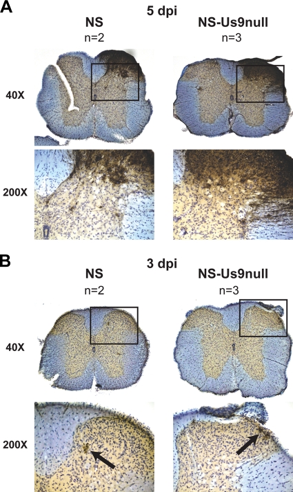
FIG. 8.
Viral spread to the spinal cord. Mice were flank inoculated with 4 × 10⁵ PFU NS or NS-Us9null. Spinal cords were dissected 5 (A) or 3 (B) dpi. Sections were stained with anti-HSV-1 and counterstained with cresyl violet. The boxed areas at ×40 magnification are shown at ×200 magnification below. Black arrows indicate viral antigen staining at 3 dpi.

