
FIG. 2.
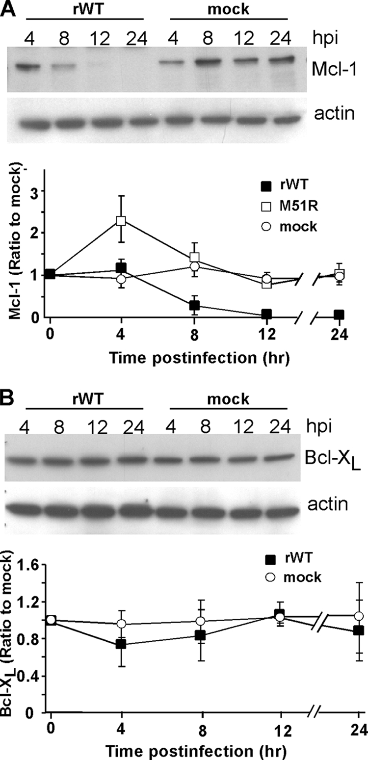
Mcl-1 levels are reduced in rwt virus-infected HeLa cells, while Bcl-XL levels remain unchanged. (A) At the indicated times postinfection with rwt virus, cell lysates were analyzed by immunoblotting with antibodies for Mcl-1. The graph shows the quantitation of Mcl-1 expression normalized to actin expression as percentages of the average ratios in mock-infected cells. The data represent the average results ± standard deviations from three experiments for cells infected with rwt virus or rM51R-M virus or mock-infected cells. (B) Cells were analyzed for Bcl-XL expression, as described for panel A.