
FIG. 3.
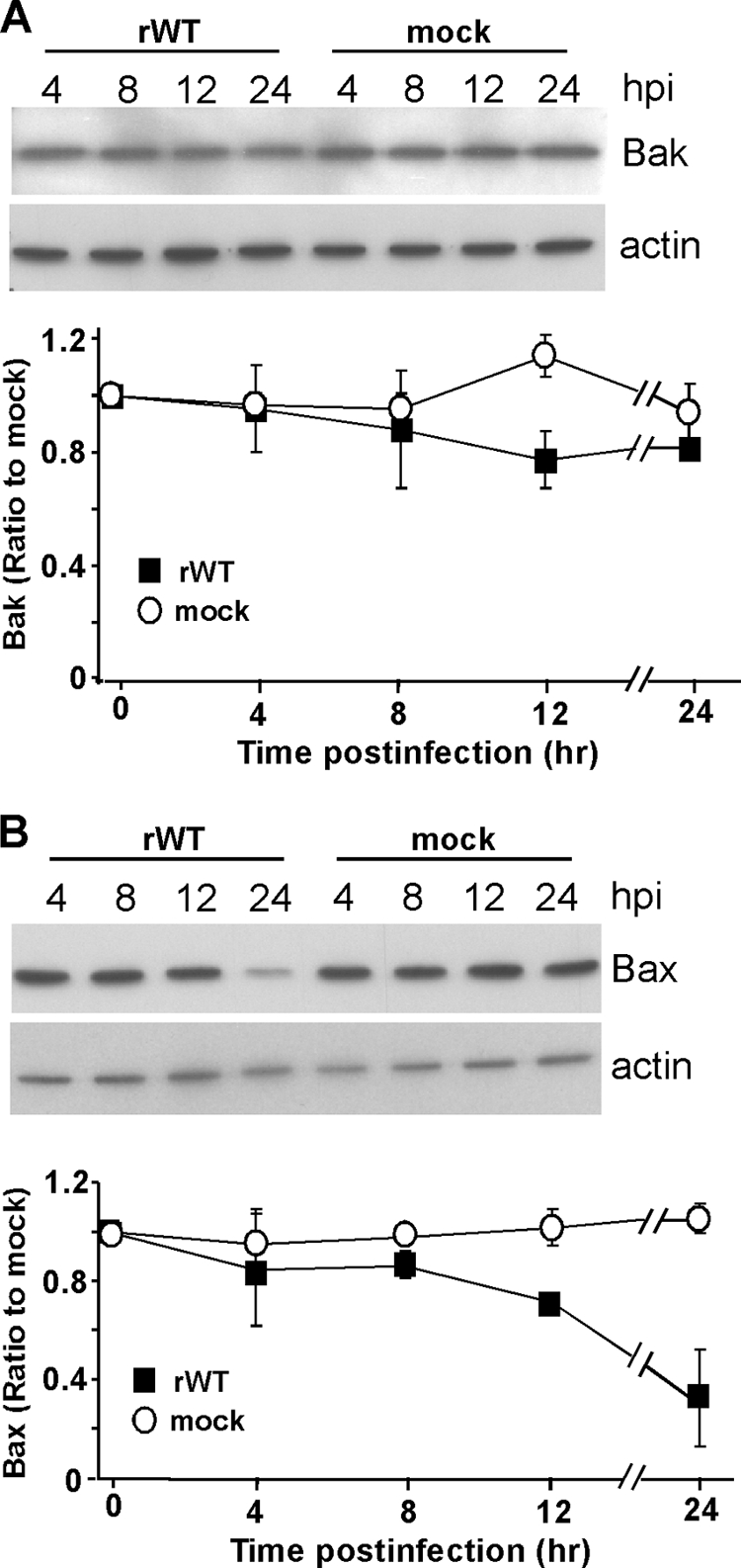
Bak levels in rwt virus-infected HeLa cells are not significantly different from those of mock-infected samples, while Bax levels decrease at later times postinfection. At the indicated times postinfection with rwt virus, cell lysates were generated and analyzed by immunoblotting with antibodies for Bak (A) and Bax (B). Representative gels are shown at the top of panels A and B, the graphs show quantitation of Bak (A) or Bax (B) protein expression normalized to that of actin protein expression, and results are shown as percentages of mock-infected samples. The data represent the average results ± standard deviations from three experiments for rwt virus-infected cells or mock-infected cells. hpi, hours postinfection.