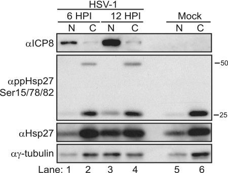
FIG. 5.
Localization of phosphorylated Hsp27 during HSV-1 infection. Nuclear (N; lanes 1, 3, and 5) and cytosolic (C; lanes 2, 4, and 6) fractions from HSV-1-infected (lanes 1 to 4) or uninfected (lanes 5 and 6) cells were collected and subjected to SDS-polyacrylamide gel electrophoresis and Western blotting. Western blots of viral (ICP8) or cellular (ppHsp27Ser15/78/82, Hsp27, and γ-tubulin) proteins are shown. The viral ICP8 protein is used as a marker for infection and an indicator of the quality of fractionation because it is a nuclear protein. “α” indicates antibody.

