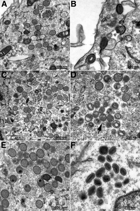
FIG. 9.
Complementation of vA17Li morphogenesis by A17 cleavage site mutants. Cells were infected with vA17Li in the absence of IPTG and transfected with plasmids expressing full-length A17 (A, B), A17G16A (C, D), or A17G185A (E, F) as described in the legend of Fig. 8. After 22 h, the cells were prepared for transmission electron microscopy. Images of fields containing IVs (A, C, E) and more mature forms (B, D, F) are shown. The arrow in panel D points to a cluster of abnormal virions.

