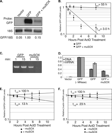
FIG. 2.
muSOX enhances mRNA degradation. (A) 293T cells were transfected with the indicated plasmids for 24 h. Total RNA was then isolated and analyzed by Northern blotting with GFP and 18S probes. Quantification (normalized to 18S levels) is shown, with the level of GFP mRNA in the absence of muSOX set to 1.0. (B) Cells were transfected as described above, and the GFP mRNA half-life was calculated by qPCR at different time points post-ActD (2 μg/ml) treatment. (C) IVT muSOX or GFP (as a negative control) was incubated for 1 or 15 min with linear plasmid DNA in degradation assay buffer at 37°C. The DNA was then extracted, resolved by agarose gel electrophoresis, and visualized by ethidium bromide staining. (D) 293T cells were transfected with empty vector or with pCDEF3-GFP alone or together with pCDEF3-muSOX. With the exception of the control (−MNase), all samples were treated with micrococcal nuclease prior to cell lysis to remove extracellular nucleic acids. Samples were then divided in half and harvested for either total cellular DNA or RNA. GFP DNA and mRNA levels were then calculated via qPCR. (E and F) Cells were transfected as described for panel A. The endogenous GAPDH (E) or β-actin (F) mRNA half-life was then calculated via quantification of Northern blots (normalized to 18S) at different time points post-ActD treatment. Errors bars show the standard error between samples. All graphs represent a compilation of at least three independent experiments. t1/2, half-life.

