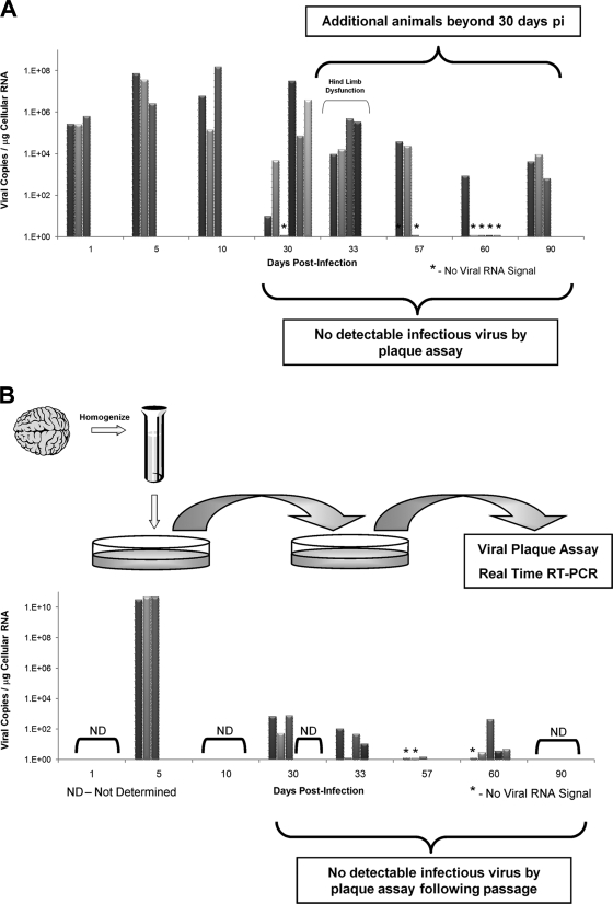
FIG. 6.
Viral RNA persistence in the CNS and in vitro passage of noncytolytic virus in tissue culture. Additional animals were infected at either 1 or 3 days postbirth with rCVB3 (2 × 10⁶ to 107 PFU i.c.), and the brains were harvested up to day 90 p.i. None of the additional mice harvested beyond day 30 p.i. had detectable levels of infectious virus, as determined by viral plaque assay. (A) In contrast, 15 of 21 animals analyzed at or after 30 days p.i. contained viral RNA, as detected by real-time RT-PCR. Of interest, one litter of pups euthanized after 33 days and suffering from hind limb dysfunction appeared to have relatively high copy numbers of viral RNA in the brain. The histopathology of one animal from this litter is depicted in Fig. 4. (B) The brain homogenates from 3 acutely infected pups and 15 infected animals (day 30 p.i. and beyond) displaying no infectious virus by plaque assay were passaged on HeLa cells. Supernatants from treated HeLa cells were isolated and plated onto fresh HeLa cells. After 1 h, supernatants were removed, and the HeLa cells were washed and placed in complete medium. After 24 h, supernatants only (no cell lysates) from the second-passage HeLa cells were analyzed by viral plaque assay and real-time RT-PCR for viral RNA. As expected, second-passage supernatants from acutely infected pups gave rise to high levels of infectious virus (as determined by virus plaque assay) and viral RNA copies. Surprisingly, low levels of viral RNA, in the absence of infectious virus, were observed in a high proportion (12 of 15) of HeLa cell supernatants from infected animals.

