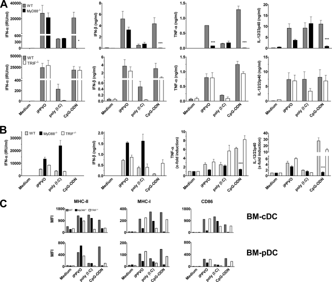
FIG. 5.
Role of intracellular adaptors MyD88 and TRIF in iPPVO-induced BMDC activation. (A) Flt3L-generated BMDC originating from wild-type (WT), MyD88−/− (upper panels), or TRIF−/− (lower panels) bone marrow were cultured in the presence of iPPVO (MOI = 5). Poly(I:C) (100 μg/ml; TRIF dependent) and CpG-ODN (1 μM; MyD88 dependent) were used as controls. Cell culture supernatants of at least duplicate cultures were harvested and analyzed by sandwich ELISA. Pooled data of two independent experiments are shown. Each column represents mean ± the SEM. (B) BM-cDC originating from wild-type (WT), MyD88−/−, or TRIF−/− bone marrow were generated in the presence of GM-CSF containing supernatant and analyzed as in panel A. Pooled data of two independent experiments (triplicate cultures) are shown. Each column represents mean ± the SEM. *, P < 0.05; ***, P < 0.001 (compared to the wild type). (C) Flt3L-generated BMDC originating from wild-type, MyD88−/−, or TRIF−/− bone marrow were cultured as in panel A. BM-pDC and BM-cDC were electronically gated and analyzed for the expression of the indicated molecules measuring the median fluorescence intensities (MFIs). The results of one of two independent experiments are shown.

