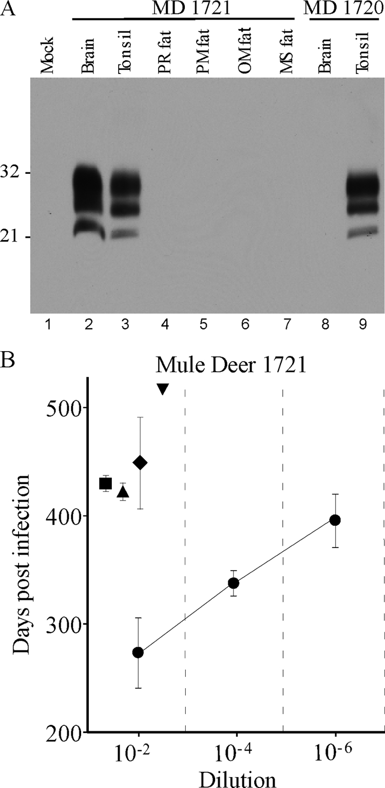
FIG. 1.

(A) Western blot of PrPres from CWD agent-infected mule deer. All samples were treated with proteinase K as described in the text. Lanes 1 and 4 to 8 were loaded with 2-mg tissue equivalents. Lanes 2, 3, and 9 were loaded with 0.5-mg tissue equivalents. Lane 1 contains uninfected deer brain, lanes 2 to 7 show tissues from mule deer (MD) 1721, and lanes 8 and 9 show tissues from mule deer 1720. The blot was probed using the anti-PrP antibody 6H4 (Prionics) by enhanced-chemiluminescence detection. Film exposure was 14 min. Numbers at the left are molecular weights (in thousands). PR, perirenal; PM, perimuscular; OM, omental; MS, mediastinal. (B) CWD incubation periods in TgDeerPrP mice following intracerebral injection of tissue homogenates from mule deer 1721. Symbols: •, brain; ▪, perirenal fat; ▴, perimuscular fat; ♦, omental fat; ▾, mediastinal fat (only one of four mice developed CWD, so there are no error bars). Note that the incubation periods found in fat at a 10−2 dilution are slightly longer than the incubation periods following inoculation of a 10−6 dilution of brain, suggesting 10,000- to 100,000-fold-lower infectivity in fat than in brain.
