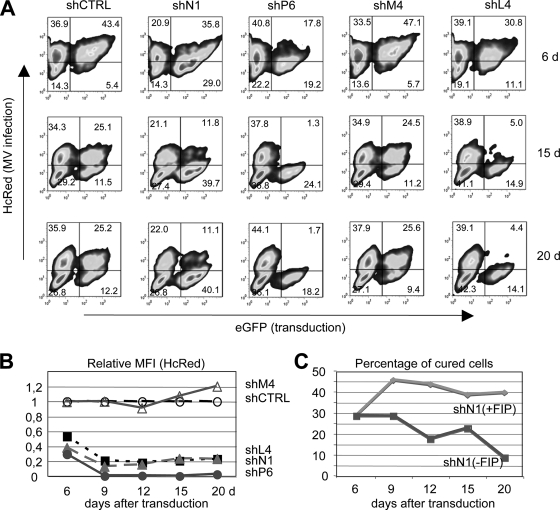
FIG. 5.
Transduction of piNT2-HcRed cells with pseudotyped lentiviral particles expressing anti-MV shRNAs. (A) Persistently infected NT2 cells were transduced with pseudotyped particles mediating the expression of control (shCTRL), N1, P6, M4, or L4 shRNAs at an MOI of 2.5 and cultivated for 6, 15, and 20 days in the presence of FIP (Z-Phe-Phe-Gly). Cell aliquots were analyzed by flow cytometry. HcRed-positive cells are infected (top left and right quadrants), EGFP-positive cells are transduced (top and bottom right quadrants), and double-positive cells are infected and transduced (top right quadrant). The relative MFIs of HcRed in transduced EGFP-positive cells (top and bottom right quadrants) are shown in relation to the control shRNA in panel B. The HcRed signal stays high after treatment of the cells with control shRNA (shCTRL, unfilled circle [set to 1]) and shRNA against M (shM, unfilled triangle), whereas shRNAs against L, N, and P (shL4, black square; shN1, gray triangle; shP6, gray circle) led to a decrease of HcRed. (C) The percentage of EGFP-positive/HcRed-negative cured cells (bottom right quadrants in panel A) is shown over time for shN-treated cells in the presence (+FIP) and absence (−FIP) of FIP.

