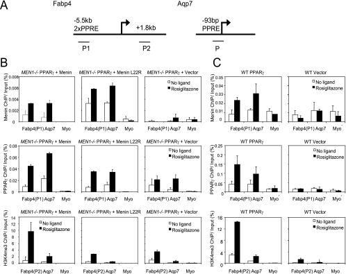
FIG. 5.
Menin recruits H3K4 trimethylation activity to PPARγ target genes. (A) Schematic representation of the Fabp4 and Aqp7 genes. Fabp4 contains two PPREs in an upstream enhancer element. For ChIP analysis, two primer pairs were used: P1 for amplifying the region of the PPREs for studying menin and PPARγ occupancy and P2 in the open reading frame to measure H3K4me3. The PPRE of Aqp7 is located close to the transcription start site; one primer pair was used for ChIP analysis. (B and C) Cell lines used for mRNA expression analysis (B) and polyclonal wild-type (WT) MEFs expressing either PPARγ or vector (C) were used. Cells were stimulated for 6 h with 1 μM rosiglitazone or DMSO. Averages of immunoprecipitated DNA, as a percentage of input, and standard errors of the means of two independent experiments are shown. (B and C, upper and middle panels) Menin and PPARγ recruitment was assessed on the PPREs of Fabp4 and Aqp7. (B and C, lower panels) H3K4me3 levels were measured in the open reading frame of Fabp4 and near the transcription start site of Aqp7. The myoglobin (Myo) gene was used as an internal negative control. (B, upper right panel) The MEN1−/− vector cell line that does not express menin serves as a negative control for the menin antibody. (C, right panels) The wild-type MEF cells that do not express exogenous PPARγ serve as a control for PPARγ in menin recruitment.

