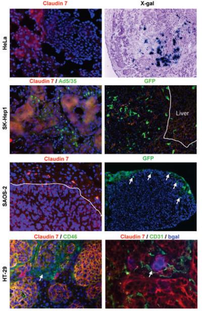
Fig.5. Correlation between epithelial and mesenchymal phenotype of liver metastases and Ad5/35 vectors transduction after intravenous injection.
Human tumor cells (HeLa, SK-Hep1, SAOS, or HT-29) were injected into the portal vein of immunodeficient mice. After liver metastases formed, mice received a tail vein injection of 2 × 10⁹ pfu of Ad5/35 vectors expressing either GFP or βGal. Tumor-bearing livers were analyzed 3 days later for claudin 7 and transgene expression. HT-29 tumor bearing liver sections were also stained for CD31. For SK-Hep1 tumors, viral particles were visualized with an anti-hexon-FITC antibody at 2 hours post-injection.

