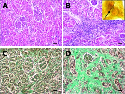
Figure 2.
A) Normal renal parenchyma from wild boar seropositive for Leptospira spp. (hematoxylin and eosin [HE] staining). B) Kidney from a seropositive wild boar, showing chronic interstitial nephritis (HE staining). Inset: silver-stained leptospire (arrow) within the tubulus epithelium of the kidney (Warthin-Starry, oil ×1,000). C) Normal renal parenchyma (Masson trichrome staining). D) Kidney with severe interstitial fibrosis (green) as a result of chronic interstitial nephritis in a wild boar seropositive for Leptospira spp. (Masson trichrome staining). Scale bars represent 50 μm.

