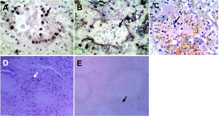
Figure 3.
Terminal deoxynucleotidyl transferase–mediated dUTP-biotin nick end-labeling staining showing numerous apoptotic alveolar epithelial cells in lung of patient B (A) and leukocytes in lung of patient A (B). C) Lung tissue from a patient with pneumonia caused by human influenza A (H5N1) virus showing apoptosis only in leukocytes. D) Spleen of patient B showing numerous apoptotic cells. E) Normal spleen tissue showing only a minimal level of apoptosis. Apoptotic cells are stained dark blue and an apoptotic cell in each panel is indicated by an arrow. Magnification ×400 in A, B, and C; ×100 in D and E.

