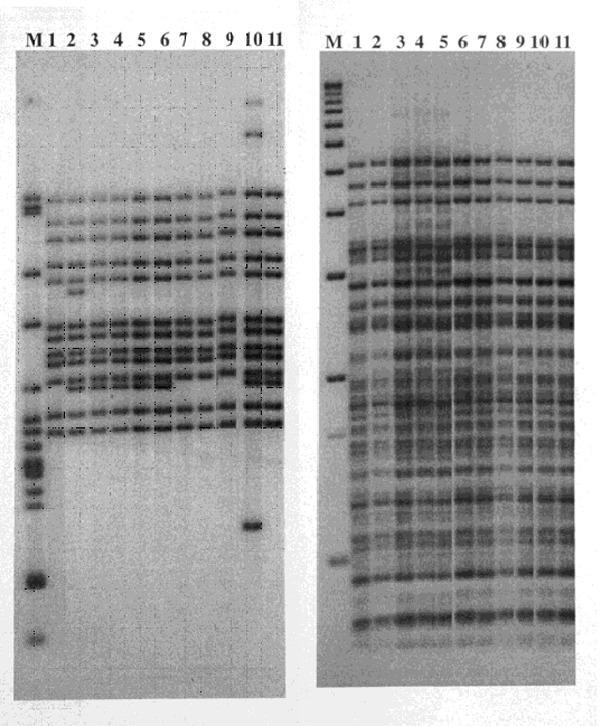
Figure 1.

Restriction fragment length polymorphism patterns of Mycobacterium tuberculosis isolates from 11 patients residing in two geographically contiguous counties, Arkansas, 1992–1998. IS6110 patterns are shown on the left and polymorphic GC-rich sequence on the right. Lane M shows M. tuberculosis strain H37Rv DNA marker (left) and 1-kb DNA ladder (right). Lane 1, isolate from patient 11; Lane 2, patient 13; Lanes 3–6, patients 4, 1, 3, and 2; Lanes 7–9, patients 10, 9, and 8; Lane 10, patient whose isolate differed by three bands and was not included in the study; and Lane 11, patient 5.
