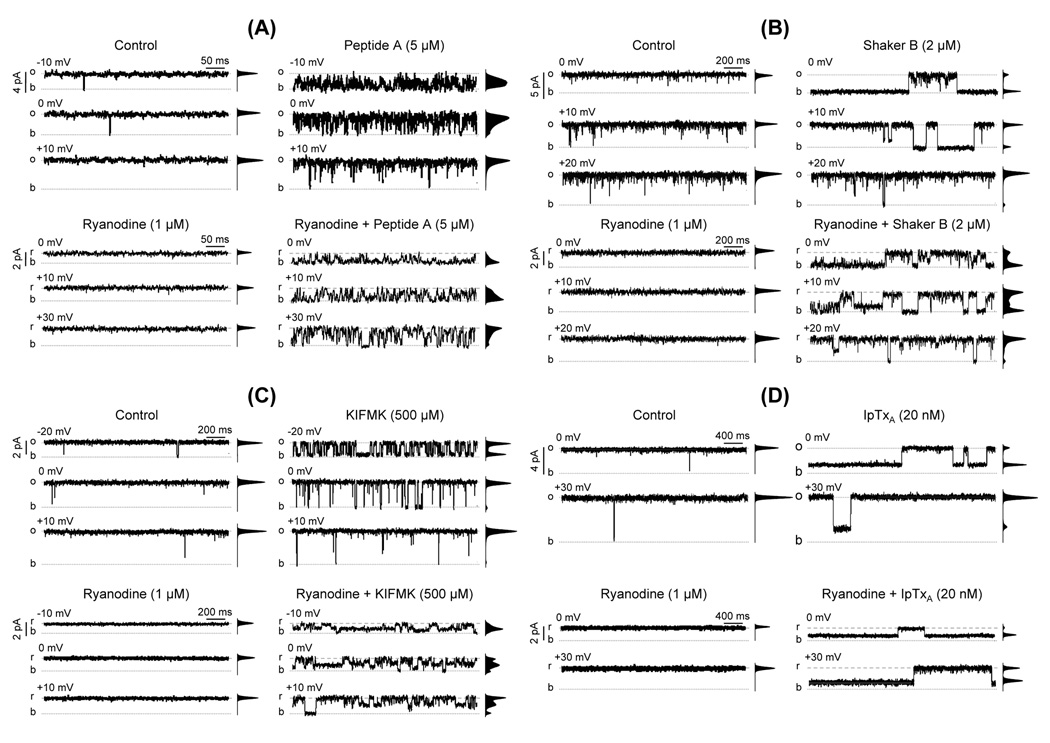
Fig. 2.
Ryanodine modifies the action of peptides and of Imperatoxin A. The figure shows single-channel recordings of single RyR2 channels activated with Ca2+/caffeine. Openings are shown as upward deflections (o = full open; b = baseline). The frequency-current amplitude histograms obtained from 4 min single-channel recordings are shown next to each trace. The figure shows the effects of (A) 5 µM Peptide A, (B) 2 µM Shaker B, (C) 500 µM KIFMK and (D) 20 nM Imperatoxin A. In all cases four panels are shown. Upper panels. Representative traces recorded at the indicated voltages in the absence of ryanodine under control conditions (upper left panels) and after addition of 5 µM Peptide A, 2 µM Shaker B, 500 µM KIFMK or 20 nM Imperatoxin A (upper right panels in A, B, C and D, respectively). Lower panels. Single RyR2 channel recordings after modification by ryanodine (2 µM). r represents full openings to the ryanodine-modified state. Traces are shown under control conditions (lower left panels) and after addition of Peptide A, Shaker B, KIFMK or Imperatoxin A (lower right panels in A, B, C and D, respectively).

