Fig. 4.
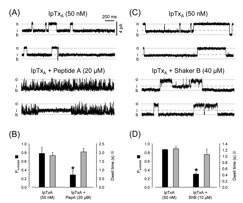
Mutual exclusion of Imperatoxin versus Peptide A or Shaker B. (A) Single-channel recordings of RyR2 with Ca2+/caffeine and IpTxA (50 nM) at Vm = +10 mV. Traces under control conditions (top) and after adding 20 µM Peptide A (bottom). (B) Probability (Psubstate; black bars) and dwell time (grey bars) of imperatoxin-induced substates before and after addition of Peptide A. * p < 0.05, n=5 paired observations. (C) RyR2 recordings in presence of Ca2+/caffeine and IpTxA (50 nM); Vm = +10 mV. Traces are shown in the absence (control; conditions (top) and presence of 40 µM Shaker B (bottom). (D) Probability (Psubstate; black bars) and dwell time (grey bars) of imperatoxin-induced substates before and after addition of 40 µM Shaker B. * p < 0.05, n=4 paired observations.

