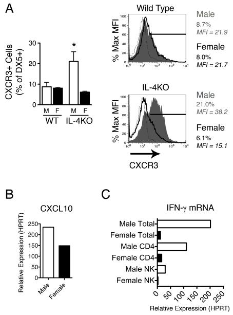
Figure 3.
Natural Killer cells derived from male IL-4KO mice express higher levels of the CXCL10 receptor (CXCR3). A) Mesenteric lymph node cells (n=4) were extracted from the MLNs of male and female WT and IL-4KO BALB/c mice at day 18 p.i. and stained for the NK marker DX5 and the chemokine receptor CXCR3. DX5+ cells were gated and the percentage of DX5+ CXCR3+ cells assessed (top panel). Plots indicate isotype control (dotted line), female (blue line) and male (red fill) MFI and percentage of DX5+ CXCR3+ cells. Representative FACs plots from each group are shown (bottom panel) B) The relative expression of CXCL10 was assessed in pooled total MLNCs (n=4) from male and female IL-4KO mice at day 28 p.i. in comparison to naïve controls via real time PCR following normalisation to HPRT. C) Expression of IFN-γ mRNA in total MLNCs and sorted CD4+ T cell and DX5+ NK cell populations was assessed in male and female IL-4KO mice at day 28 p.i. * - p≤0.05

