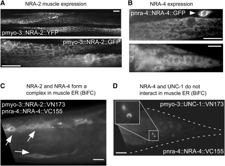
Figure 2.
NRA-2 and NRA-4 are expressed in the ER and interact in a complex. (A) NRA-2∷YFP (upper panel, single confocal plane) or NRA-2∷GFP (lower panel, epifluorescence) were expressed from the muscle-specific pmyo-3 promoter. Reticular expression, reminiscent of the ER was found. (B) NRA-4∷GFP was expressed from the endogenous pnra-4 promoter. Intracellular, reticular expression was observed in muscle cells (upper panel) and neurons (arrowhead), and in other tissues (lower panel: muscles, neurons and hypodermal cells in the tail). (C) NRA-2 and NRA-4 form a complex, as shown by bimolecular fluorescence complementation (BiFC). NRA-2 was fused to the VN173 fragment of Venus, and NRA-4 to the VC155 fragment. Fluorescence was restored in muscle ER (arrows point to muscle cell nuclei surrounded by ER), in which the two proteins were co-expressed. (D) NRA-4∷VC155 does not interact in the ER with the stomatin UNC-1∷VN173, expressed in muscle (a gift by ZW Wang). Occasionally, vesicular fluorescent structures were observed, possibly representing lysosomes in which the fusion proteins are degraded and in whose membranes their cytosolic tails (and Venus fragments) accumulate. Size bars are 10 μm.

