Figure 3.
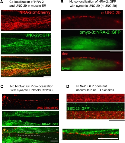
NRA-2 co-localizes with L-AChR subunits in the ER, but not at synapses. (A) NRA-2∷mCherry (expressed from the pmyo-3 promoter) was co-expressed with the L-AChR subunit UNC-29∷GFP (expressed from punc-29) and co-localization was observed by confocal microscopy (single confocal plane of midbody muscle cells). (B) Endogenous UNC-29 protein was immunolabelled with specific antibodies in animals expressing NRA-2∷GFP in muscles (GFP fluorescence was preserved during fixation). Dorsal nerve cord (dnc) and adjacent muscle cells (bwm) are shown near the pharyngeal terminal bulb. No co-localization of NRA-2∷GFP and UNC-29 was apparent. (C) NRA-2∷GFP was co-expressed in muscle with epitope-tagged UNC-38∷3xMYC (expressed from punc-38). UNC-38, exposing the MYC tag on the cell surface, was labelled with Cy3-conjugated anti-MYC antibodies injected into the body cavity. The ventral nerve cord was imaged by confocal microscopy (single focal plane), showing punctate cell-surface L-AChR clusters that contain UNC-38. NRA-2∷GFP is adjacent to L-AChR clusters, but not co-localizing with them (inset: enlarged region). (D) SEC-23∷GFP, a COPII coat component that labels ER exit sites, and NRA-2∷mCherry were co-expressed in muscle and imaged by confocal microscopy. Puncta of SEC-23 accumulation contained also NRA-2; however, NRA-2 did not accumulate at these sites. Z-stack of confocal sections. Size bars are 10 μm.

