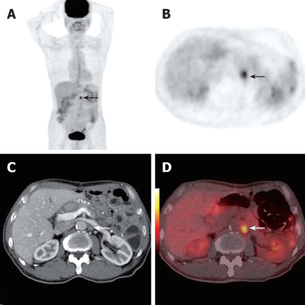
Figure 2.
A 75-year-old asymptomatic man who had gastric cancer resection 1 year previously underwent PET/CT as part of routine post-operative surveillance. Whole body PET projection image and axial PET image showed focal hypermetabolic activity in the abdomen (black arrow, A and B). Axial contrast CT detected a small lymph node at the same position (white arrow, C). PET/CT fusion images showed a focus of highly metabolic metastasis in retroperitoneal lymph node (white arrow, D).This was later verified by follow up. The case illustrated the value of early discovery by PET/CT in asymptomatic patients after surgery.

