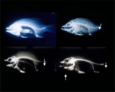
Figure 5.
Different Types of Skeletal Deformities in Tilapia.
Upper left: X-ray of tilapia collected from Lin-Bien River in October, 1994. Arrow shows vertebral segments close to tail are fused, calcified and deformed. Lower left: X-ray of tilapia collected from TKS station of Tongkong River in October, 1994. Note fusion of vertebrae near the tail (arrow). Upper right: X-ray of tilapia collected from the mouth of the Tongkong River in May, 1995. Note lordosis near the tail (arrow). There was also muscular atrophy in the tail area as well. Lower right: X-ray of tilapia collected from Kao-Ping River in October 1994. Arrow shows heavy calcification in the lower jaw, and fusion of vertebrae near the tail.

