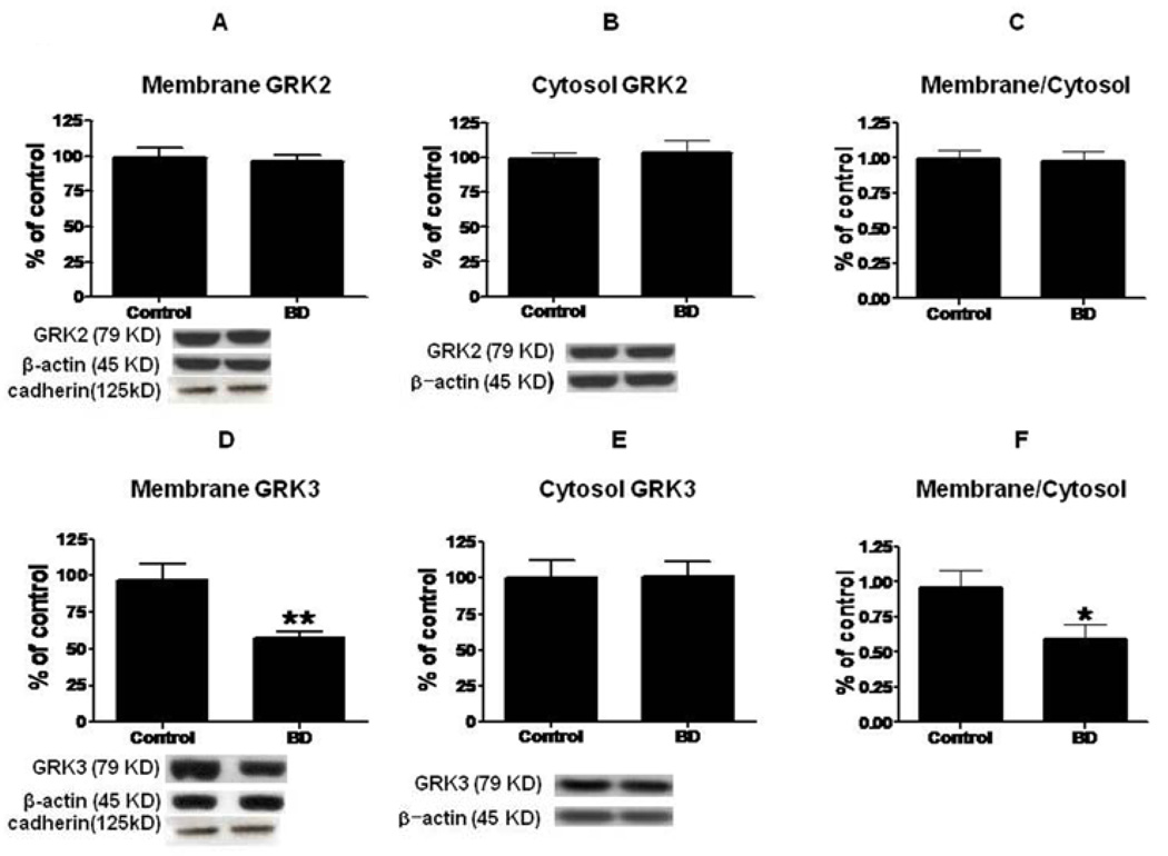
Figure 2.
Representative immunoblots of GRK2 and GRK3 protein levels in membrane (A, D) and cytosol (B, E) in frontal cortex of controls (n = 10) and BD patients (n = 10). Data are ratios of optical density of GRK2 and GRK3 to β-actin, expressed as percent of control, and compared using a two-tailed, unpaired t-test (mean ± SEM, *p < 0.05, **p < 0.01). Bar graphs of membrane to cytosol ratios of GRK2 (C) and GRK3 (F) in frontal cortex of controls and BD patients (Mean ± SEM, *p < 0.05).

