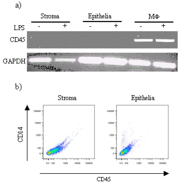
Figure 3.

Analysis of CD45 expression by stromal and epithelial cells. A) Cells were stimulated with LPS for 24h, harvested and RNA was isolated as described and the resulting cDNA analysed for the presence of CD45 transcripts using the indicated primer pairs. cDNA from bovine MΦ were used for comparative reason. B) stromal and epithelial cells were stained with antibodies to bovine CD45 and surface expression of these molecules analysed by flow cytometry. A representative staining result is shown (n=3).
