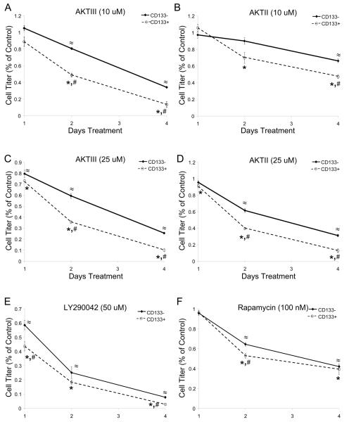
Figure 3. Targeting Akt Preferentially Decreases CD133+ Brain Tumor Cell Growth Over Time.
CD133+ and CD133− cells isolated from a T3359 glioblastoma patient specimen passaged short-term in immunocompromised mice were plated in Neurobasal media with EGF and FGF, allowed to recover overnight, and then treated with 10 uM AktIII (A), 10 uM AktII (B), 25 uM AktIII (C), 25 uM AktII (D), 50 uM LY290042 (E), and 100 nM rapamycin (F) inhibitors. Cell growth was measured on the indicated days after inhibitor treatment began using the Cell Titer Glo assay (Promega) according to the manufacturer's instructions. The data for each time point were standardized to the DMSO treated controls for the same cell type on each day. *, p<0.05 with t-test comparison of inhibitor treated CD133+ cells to DMSO treated control CD133+ cells on the same day; ≈, p<0.01 with t-test comparison of inhibitor treated CD133− cells to DMSO treated control CD133− cells on the same day; #, p<0.05 with t-test comparison of CD133+ cells to similarly treated CD133− cells on the same day.

