Figure 2.
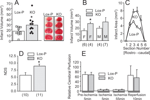
Neuronal PPARγ deficiency aggravates cerebral ischemic damage. N-PPARγ-KO (KO) and control (Lox-P) mice were subjected to reversible 60 min MCA/CCA occlusion. A, The infarct volumes representing composite of all the control (white filling) and KO (gray filling) mice (individual mice are indicated as squares or diamonds within each group), (B) segregated by gender (M, male; F, female; number below each bar indicates the sample size), *p < 0.05, and infarct areas (C) at six rostrocaudal plains were determined from morphometric analyses of TTC-stained brain sections at 3 d after the onset of ischemia. Representative photographs of TTC-stained brain sections demarcate infarcts in control (Lox-P) and N-PPARγ-KO (KO) mice (included in A). Infarct volume in all groups was established at 3 d after ischemia. D, Bar graph illustrating the NDS as measured at 3 d after ischemia using a battery of behavioral tests. Number below each bar indicates the sample size. *p < 0.05. E, Bar graph illustrating cerebral perfusion values in the peri-ischemic areas of control (n = 10) and N-PPARγ-KO mice (n = 11) at four time points representing 5 min before ischemia (Preischemia; baseline), 5 min and 55 min after induction of ischemia, and 10 min after reversal of the occlusion (reperfusion) measured using Laser Doppler flowmetry. The results are presented as mean ± SEM.

