
Fig. 7.
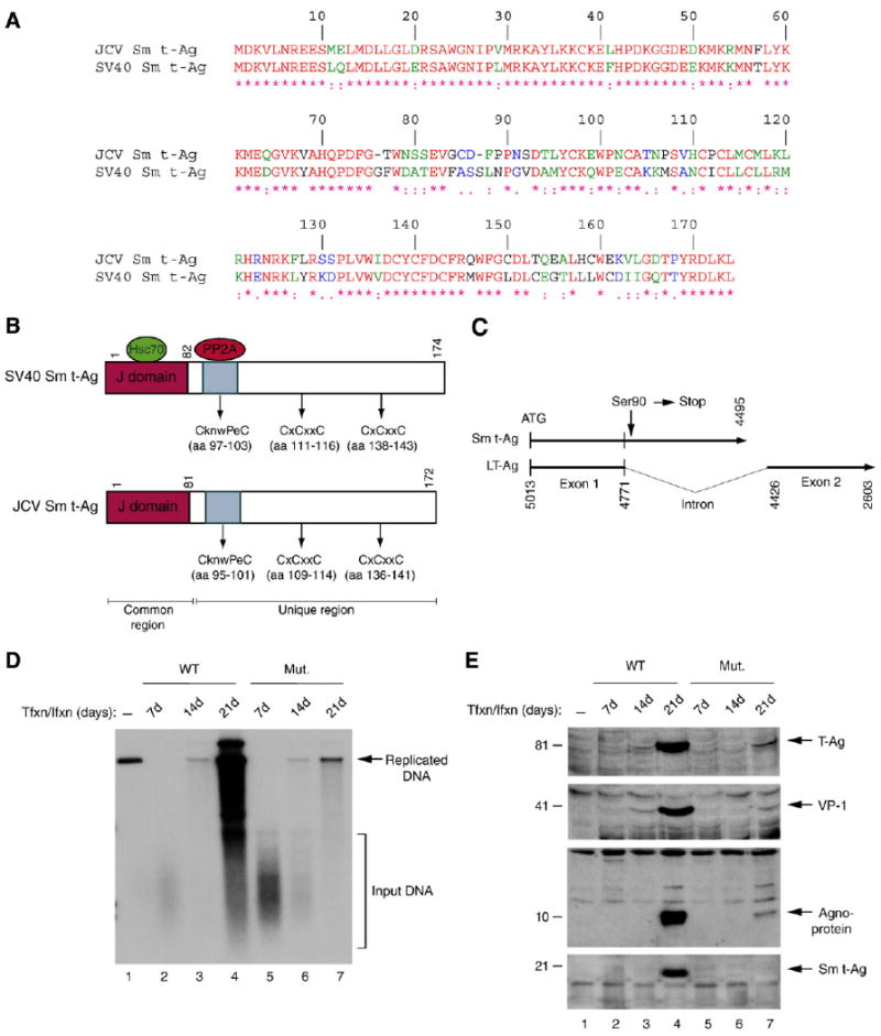
Viral replication and gene expression were substantially down-regulated in the absence of full-length Sm t-Ag expression. (A) Amino acid alignment of JCV Sm t-Ag and SV40 Sm t-Ag. (B) Schematic comparison of SV40 Sm t-Ag regions with that of JCV Sm t-Ag. The position of J domains, PP2A binding domains and cystein clusters are indicated. (C) Schematic representation of JCV Mad-1 early coding region, where Ser90 (TCT) of Sm t-Ag was converted into a stop codon as described in Materials and methods. Numbering is according to JCV Mad-1 strain (GenBank # NC_001699, formerly J02226). (D) Dpn I assay. Primary human fetal glial cells (PHFG) (4 million cells/75 cm2 flask) were transfected/infected either with WT (5 μg) or Mut genome (5 μg) by lipofectin method. At 7d, 14d and 21d posttransfection, low molecular weight DNA was isolated (Ziegler et al., 2004) and analyzed by Dpn I assay (Hirt, 1976). (E) In parallel, either nuclear or cytoplasmic extracts were prepared from transfectants and analyzed by Western blotting using anti-LT-Ag (PAb-2000), anti-VP-1 (PAB597, a gift from Dr. W. Atwood), anti-agnoprotein and anti-Sm t-Ag antibodies as indicated.