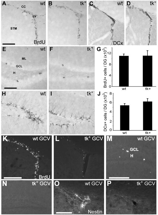
Figure 2.
A–J: Immunolabeling for BrdU (A–D) and DCx (F–I) shows robust cell proliferation and production of immature neurons in both the SVZ (A, B, F, G) and DG (C, D, H, I) of untreated, 3 month old wild-type (wt) and nestin-tk+ (tk+) mice. No significant difference in stereological estimates of the number of BrdU (E) or DCx (J) immunolabeled cells in the DG was observed between untreated wt or tk+ mice. K–P: Intraperitoneal injection of eGCV for 14 days dramatically reduces proliferation, as indicated by labeling for pulse-administered BrdU, in the SVZ (L) and SGZ (N) of tk+ mice relative to wt controls (K, M). BrdU was injected 2 hours prior to perfusion. The population of nestin positive cells in the SVZ is also depleted in tk+ mice (P) compared to controls (O). LV, lateral ventricle; STM, striatum; CC, corpus callosum; GCL, granule cell layer; H, hilus; ML, molecular layer. Scale bar = 200 μm in A, B, F, G, K–N and 100 μm in C, D, O, P.

