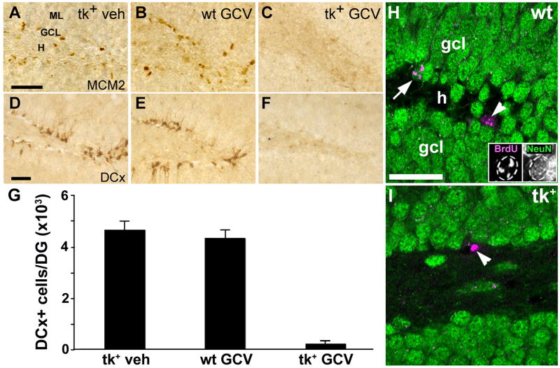
Figure 4.
ICV infusion of GCV for 28 days dramatically reduces both cell proliferation and the number of immature neurons in the DG of nestin-tk+ (tk+) mice. A–C: While the SGZ of both vehicle-treated nestin-tk+ (tk+) and GCV treated wild-type (wt) mice contains numerous mitotically active cells expressing Mcm2 (A, B), few cells label for this cell cycle marker in the SGZ of tk+ mice treated with 28 days of GCV (C). D–F: Immature neurons immunolabeled for DCx are absent from the DG of tk+ mice treated with GCV (F), but are not depleted in vehicle-treated tk+ (D) or GCV-treated wt (E) controls. G: Quantitative analysis shows that DCx-immunoreactive cell numbers are 95% lower in GCV-treated tk+ mice than in either control group (p < 0.01). H, I: Representative confocal images of sections double-labeled for BrdU (magenta) and NeuN (green) show both double- (arrow) and single-labeled (arrowhead) cells in the granule cell layer (GCL) of a GCV-treated wt control (H), but only a cell single-labeled for BrdU in a tk+ animal after 28 days of GCV treatment (arrowhead in I). Non-merged images of the double-labeled cell (arrow) are shown as insets in H. BrdU was given 16–18 days earlier (days 10–12 of GCV infusion). H, hilus; ML, molecular layer. Scale bars = 50μm.

